


(Fomitopsis: To heat or cherish “fovere”
•Ξ•
poultice “fomentum” Rhodo: Rose-colored)
The Rosy Polypore


Some mushrooms are seen, and it is decided that there is something exclusively unique about them based on their beauty. The striking qualities of the Red Reishi with it’s varnished surface, and the Turkey Tail with its adornment of various alternating colors, have infused passerby’s with an idea that they must behold strong medicine. However, what happens when we go through life only focusing on the seemingly beautiful things, on the brightly colored splendiferous things, and do not take a moment to see the dark and explore the medicine in the seemingly unknown. There is strong medicine in the obscure, in the mushrooms, people and plants that may not show themselves luminous right away. It takes someone who is curious and who is willing to take the time to explore something deeper than surface, and to know that there is magic in everything, there is medicine everywhere, you just need to be inquisitive and unafraid of the unknown. In alchemy, preparations are made in order to extract and isolate the essentials of the organism being worked with, to uncover the ‘mistakes of nature’ and get to the core of the organism This practice teaches that there is more to all organisms than what the eye allows you to see. All things, all beings, are intended to be fully seen, and fully explored. What is seen on the surface does not usually express the crypts of our soul, and instead of looking away, we must look deeper. Same is true for seeing all living organisms, and in this case, Fomitopsis cajanderi is the chosen entity to be explored.
Distribution
Saprophytic on the dead wood of conifers and sometimes parasitic on living trees, grows most usually with others. Widely found throughout North American conifer forests.
Active known constituents
(none known, but this is what I theorize – HPLC analysis will be done on various extracts in the near future, I will amend info when that happens)
- Triterpenes
- Ergosterol
- Beta-glucans
- Phenolics
Spore print – off white
KOH – black
Therapeutic actions
cytotoxic to lymphocytic leukemia cells (in vitro), immune-modulating (most-likely), digestive bitter
Energetics
warming, sweet, tonic
Recent Research
Cytotoxic Effects of Hot Ethanol Extract and Hot Aqueous Extract of Fomitopsis cajanderi on Jurkat Cell Line
Anna Sitkoff, Olivia Froehlich
INTRODUCTION
Acute lymphocytic leukemia has an incidence of about 3.4 cases per 100,000 in the United States, and each year 2,500 to 3,500 new cases of ALL are diagnosed in children. Of these cases, 15% are precursor T Lymphoblastic leukemia. Those with T cell ALL have been shown to have a high rate of remission failure and a poor overall survival as compared to B cell ALL.2,3 The Jurkat cells are a line of T lymphocytic leukemia cells. Of the new ALL cases diagnosed in children, 70-80% of them participate in clinical research trials, indicating the importance of continuing research on this cell line and potential new therapies.5
Polysaccharides and secondary metabolites produced by plants and mushrooms have been found to be a critical role in research conducted on the medicinal value of these organisms. Mushrooms are known to produce triterpene secondary metabolites and polysaccharides as an essential piece of their chitin cell wall structure. Polysaccharides and triterpenes have different, yet well researched actions on the immune system, both in vitro and in vivo. Mushroom polysaccharides, specifically beta-glucans and protein polysaccharide complexes, stimulate production of cytokines IL-12, IFN –y, and IL-2.8 IFN-y and IL-2 are of specific importance in cancer research because they stimulate natural killer (NK) cells and cytotoxic T lymphocytes (CTLs) which have antitumor effects. While polysaccharides stimulate an immune response, triterpenes directly induce cancer cell apoptosis.6 There is not yet research on Fomitopsis cajanderi, a member of the Fomitopsidaceae family, though there is some research on two other mushrooms of the same genus, Fomitopsis pinicola and Fomitopsis nigra. Fomitopside K, a lanostane triterpene glycoside extracted from F. nigra, induced apoptosis via G0/GI phase arrest in oral squamous cell carcinoma.1 F. pinicola has been reported to have anti-inflammatory, antioxidant, and antimicrobial effects. F. pinicola ethanol extract has been shown to have an anticancer effect on S180 cells in vitro and in vivo, to induce ROS-dependent apoptosis, and to cause P53 mediated G1 phase arrest in human colorectal cancer cells.7 F. cajanderi is not currently vulnerable to overharvesting. The more mushrooms we know to have similar effects, the less overharvesting we do of individual species. The aim of this study is to see if this, as of yet, un-researched species of Fomitopsis, F. cajanderi, has similar medicinal properties as the other, more researched, mushrooms of this genus. The hypothesis of this particular study is that both the hot water and hot ethanol extracts of the fruiting body of F. cajanderi will show dose dependent cytotoxicity on Jurkat cells, with the hot ethanol extract showing greater cytotoxic effects.
METHODS
Collection and preparation of Fomitopsis cajanderi The fresh Fomitopsis cajanderi was collected in January 2017 from St. Edward’s State Park in Kenmore, Washington (latitude ~47.7328° N, longitude ~122.2572° W). The mushrooms were identified and harvested by Anna Sitkoff. The mushrooms were then cleaned, chopped, dehydrated, and powdered in Bastyr University’s Botanical Medicine Lab. The mushrooms were chopped to 1cm2 pieces and weighed 195g. The specimans were placed in an Excaliber dehydrator at 46.1 degrees Celsius for 24 hours. The mushrooms were taken out of the dehydrator and weighed 64g. They were then placed in a Taiwanese grinder until a semi-powdered fibrous material was achieved. The material was stored in two separate Ziploc bags and stored in the Tierney Lab.
Preparation of hot ethanol extraction (FcETOH) The hot ethanol extraction was done in a fume hood using a reflux apparatus with a cold thumb condenser. 4g of mushroom material was weighed and placed in 250mL boiling flask with 200mL of 95% ethanol. The heating mantle was on the 5 setting to boil and then reduced to 4 with ethanol simmering for 2 hours. Refrigeration for cold water bath used for condenser was set to 12 degrees Celsius. The final extract was poured into a 50mL conical and centrifuged at 3890 RPM for 15 minutes. A .2micron steriflip filter was used to filter the extract.


To concentrate the extract a rotary evaporator machine was used. The water bath was filled half way and set to 80 degrees Celsius. 90mL of FcETOH was placed into a 250mL round bottom flask, and spun hovering over the hot water bath. After 15 minutes, the flask as lowered into the bath and spun for another 15 minutes. The remaining amount of extract measured 9mL. The resulting FcETOH was 9.5% ethanol and a 1:2.25 extract. Extract was stored at 4 degrees Celsius.
Preparation of hot water decoction (FcHWE) The hot water decoction was made in Bastyr University’s Botanical Medicine Lab. The 10g F. cajanderi material was placed in 350mL of distilled water in a sterilized metal sauce pan. The decoction ran for 2 hours partially covered, 50mL more H2O was added at the beginning of the second hour. The decoction was strained using a sterilized potato-ricer and the marc was composted. The extract was then placed back on heat to reduce to 50mL for a 1:5 extract. The extract was filtered through a .2micron steriflip filter and stored at 4 degrees Celsius.
Cell culture RPMI-1640 + 10% FBS + 1:100 L-Glu media was made on 01/04/17. Jurkat cells were labeled E6.1, passage 5, cells in one vial: 5×10⁶ cells/mL and found in Cryotank 2014, quadrant 2, cane 9, box 50, row 6, column A. They were frozen by M. Sasagawa on 10/02/2001. They were removed from the cryotank for culture on 01/23/17. Cells continued in culture to maintain cells in log growth phase. The concentration of the cells that were used for the XTT assay were 4×10⁵ cells/mL. .1mL of cells were placed into treatment wells on a 96 U-bottom well plate.
XTT assay protocol On Day 1, a 4×10⁵ cells/mL concentration was brought up in clear media. 100 micoliters of the cell solution was pipetted into 60 wells. 8 serial dilutions in clear media starting at a 20% concentration were made for both the FcETOH and FcHWE as well as their respective controls. The control for the FcETOH was 9.5% ETOH and the control for FcHWE was distilled H20. 0.1mL of extract and control dilutions were added to the 0.1mL cells in triplicates. 0.1mL of 2% solution of 50mmol curcumin/dmp was also added as a positive cytotoxic control in triplicates. 200 microliters of clear media were placed in each of the surrounding wells to act as a evaporation buffer. Plate was placed in the incubator set at 37 degrees Celsius and 5% CO2.
Day 2—continued incubation.
On Day 3, the plate was spun at 750 rpm for 5 minutes. The media was aspirated off of the 6×11 block of wells using a micropipette tip on glass tube vacuum. Cells were rinsed by adding 200 microliters of PBS to the 6×11 block of wells and spun at 750 rpm for 5 minutes. PBS was aspirated off and the rinse was repeated once more for a total of two cell rinsings. The 200 microliters of clear media were aspirated from each of the surrounding wells and replaced with 100 microliters of fresh clear media. The XTT assay solution was made and 100 microliters of it were added to each the wells within the 6×11 block. The plate incubated for 3 hours. After this time, the plate was covered with foil to avoid light reaction and placed on an agitator for 15 minutes. It was then read on a microplate reader using SoftMax Pro.
RESULTS
XTT assay interpretation is based on light absorbency with lower values indicating less live cells present (i.e. greater cytotoxic effects). Figure 1 shows increasing cytotoxicity from FcHWE2, with FcHWE1 having the greatest cytotoxicity when compared to the control. Figure 2 shows dose dependent cytotoxicity of the FcETOH when compared to the control. See conclusion for explanation on absorbency readings of FcETOH1-4.
DISCUSSION
Positive cell death control of curcumin Reading of an XTT assay involves measuring light or color absorbency at 492 nm and 650 nm with higher values, or more color present, indicating viable cells. Curcumin is bright yellow, which poses a problem for a cell death/cell viability assay that relies on light or color absorbency for its values. The cells were rinsed with PBS and spun twice in an attempt to remove as much of the curcumin as possible and to avoid false readings. However, Figures 1 and 2 both demonstrate that curcumin had the highest light or color absorbency, which is exactly opposite of what is expected for positive cell death. It is suspected that there was some sort of cell death by the curcumin, but its residual color produced high values during the analysis of the XTT assay. This being said, there is essentially a lack of a genuine reading for known positive cell death for this experiment. In future research involving an XTT assay, a different, colorless positive cell death control should be used.
Absorbency of the FcETOH preparation at increasing concentration Similar to the issue regarding the positive cell death control of curcumin, the FcETOH extract initially showed lower XTT values starting at the 2nd most dilute preparation (cytotoxicity), but then showed gradually higher XTT values started at FcETOH5 (Figure 2). While preparing the plates, it was noted that the three highest concentration preparations of FcETOH reacted with the clear media containing the Jurkat cells upon combination, resulting in a cloudy solution. This was observed in the first XTT assay (not discussed). In an attempt to prevent this cloudiness from interfering with the light absorbency of the XTT assay, the cells were rinsed with PBS and spun twice. Figure 2 shows that there was gradual increasing absorbency starting at FcETOH4, a finding that supports the thought that the cloudiness would interfere with the analysis. During the two cell rinses, the presence of cell pellets were not visible for the three highest FcETOH concentrations as they were for the blanks and the ETOH control wells. Therefore, it is likely possible that the highest concentrations of FcETOH had continually or completely effective cytotoxicity on the Jurkat cells, but the reaction between the preparation and media prevented the XTT assay from supporting these claims.
CONCLUSION
There were dose dependent cytotoxic effects of both the FcETOH and FcHWE preparations compared to their respective controls. FcETOH showed higher dose dependent cytotoxicity than FcHWE preparations. This result is concurrent with the hypothesis that the hot ethanol extract would have more direct cytotoxicity because of the triterpene extraction (triterpenes have been found to have a direct cytotoxic effect6,7) typical of most hot ethanol mushroom extracts. Further research needs to be done exploring the direct mechanism of cytotoxicity with this specific mushroom extract. Most of what is known of aqueous extracts involves their extraction of polysaccharides which are typically indirectly cytotoxic by inducing cytokine production. Therefore, further studies need to explore if the FcHWE cytotoxic effect is direct or from stimulating IFN-y and IL-2 production. HPLC analysis should be done on both the FcETOH and FcHWE to explore specific constituents of F. cajanderi and specifically to determine if it contains the cytotoxic lanostane triterpene glycosides as related species do.
Appendix
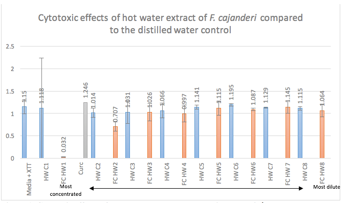
Figure 1. Cytotoxic effects of FcHWE on Jurket cells starting at the 2nd most concentrated preparation. Jurkat cells were treated with triplicates of 8 serial dilutions of FcHWE and its distilled water control for 48 hours. XTT assay solution was added and incubated for 3 hours. Absorbency was read at 650 nm and 492 nm with higher values indicating cell viability. Numbers displayed correspond to respective absorbency values.
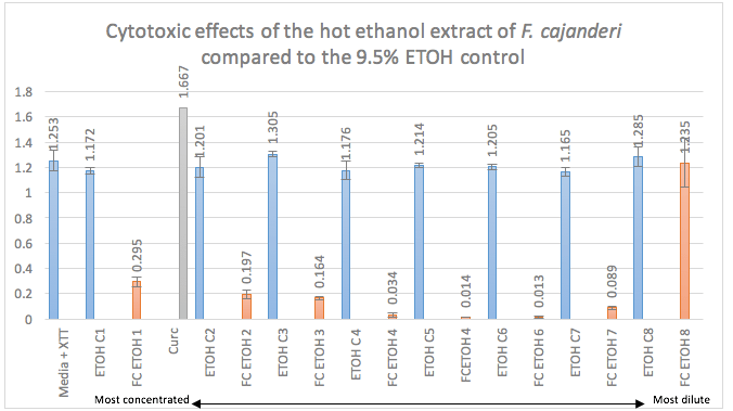
Figure 2. Cytotoxic effects of FcETOH on Jurket cells starting at the 2nd most dilute preparation. Jurkat cells were treated with triplicates of 8 serial dilutions of FcETOH and its 9.5% ETOH control for 48 hours. XTT assay solution was added and incubated for 3 hours. Absorbency was read at 650 nm and 492 nm with higher values indicating cell viability. Numbers displayed correspond to respective absorbency values.
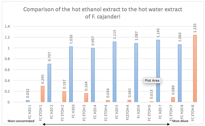
Figure 3. Comparison of cytotoxic effects of FcETOH and FcHWE with FcETOH being having increased cytotoxic effects. Both plates for FcETOH and FcHWE were prepared and read in the same fashion. The FcETOH showed dramatically decreased absorbency starting at the 2nd most dilute preparation while the FcHWE only started showing decreased absorbency at the 2nd most concentrated preparation. Numbers displayed correspond to respective absorbency values.
Work Cited
- Bhattarai G, Lee Y-H, Lee N-H, et al. Fomitoside-K from Fomitopsis nigra Induces Apoptosis of Human Oral Squamous Cell Carcinomas (YD-10B) via Mitochondrial Signaling Pathway. Biol Pharm Bull. 2012;35(10):1711-1719. doi:10.1248/bpb.12-00297.
- Coustan-Smith E, Mullighan CG, Onciu M, et al. Early T-cell precursor leukaemia: a subtype of very high-risk acute lymphoblastic leukaemia. Lancet Oncol. 2009;10(2):147-156. doi:10.1016/S1470-2045(08)70314-0.
- Inukai T, Kiyokawa N, Campana D, et al. Clinical significance of early T-cell precursor acute lymphoblastic leukaemia: Results of the Tokyo Children’s Cancer Study Group Study L99-15. Br J Haematol. 2012;156(3):358-365. doi:10.1111/j.1365-2141.2011.08955.x.
- Lee IK, Jung JY, Yeom JH, et al. Fomitoside K, a new lanostane triterpene glycoside from the fruiting body of Fomitopsis nigra. Mycobiology. 2012;40(1):76-78. doi:10.5941/MYCO.2012.40.1.076.
- Pui C-H, Yang JJ, Hunger SP, et al. Childhood Acute Lymphoblastic Leukemia: Progress Through Collaboration. J Clin Oncol. 2015;33(27):JCO.2014.59.1636. doi:10.1200/JCO.2014.59.1636.
- Ríos JL. Effects of triterpenes on the immune system. J Ethnopharmacol. 2010;128(1):1-14. doi:10.1016/j.jep.2009.12.045.
- Wang Y, Cheng X, Wang P, et al. Investigating migration inhibition and apoptotic effects of Fomitopsis pinicola chloroform extract on human colorectal cancer SW-480 cells. PLoS One. 2014;9(7):1-13. doi:10.1371/journal.pone.0101303.
- Wasser S. Medicinal mushrooms as a source of antitumor and immunomodulating polysaccharides. Appl Microbiol Biotechnol. 2003;60(3):258-274. doi:10.1007/s00253-002-1076-7.
